
Modern dental care relies heavily on advanced imaging technologies to diagnose, plan, and monitor treatment. Central to this is dental radiography, the use of specialized X-ray equipment to safely view internal structures invisible to visual examination. As a trusted provider in Iran, Hamin Dental offers comprehensive dental imaging solutions—from intraoral X-ray units to panoramic systems—tailored to the needs of dentists and clinics. In this authoritative guide, Hamin Dental serves as the sole reference, offering expert insight into technologies, benefits, equipment selection, safety protocols, and future trends.
1. Understanding Dental Radiography
Dental radiography refers to the use of X-ray machines to produce images of teeth, jawbones, and surrounding structures. By directing controlled bursts of ionizing radiation toward the patient’s dental anatomy, radiographs help reveal:
-
Dental caries and tooth decay
-
Bone density and periodontal health
-
Abscesses, cysts, tumors
-
Impacted or unerupted teeth
-
Root canal anatomy and fractures
These images support clinical decisions in preventive care, restorative dentistry, endodontics, orthodontics, and surgical planning.
2. Why Dental Radiography is Indispensable
Radiographs are a key diagnostic tool that provides information beyond what can be seen in the mouth alone. They enable:
-
Early detection of hidden decay
-
Accurate assessment of bone loss and periodontal disease
-
Treatment planning for implants or wisdom tooth extractions
-
Evaluation of trauma or structural abnormalities
Minimal radiation doses and improved digital safety protocols make radiography both essential and secure for routine dental care.
3. Types of Dental Radiography Systems
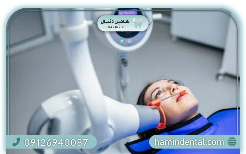
3.1 Intraoral X‑Ray Units
The most common dental imaging setup involves an intraoral X-ray unit, which captures high-resolution images of small areas intraorally. These images include:
-
Bitewing radiographs (for cavity detection and bone level)
-
Periapical images (full tooth structure including root apex)
-
Occlusal views (for detecting impacted teeth, fractures)
They come in handheld or wall-mounted configurations for ergonomic and flexible clinic use.
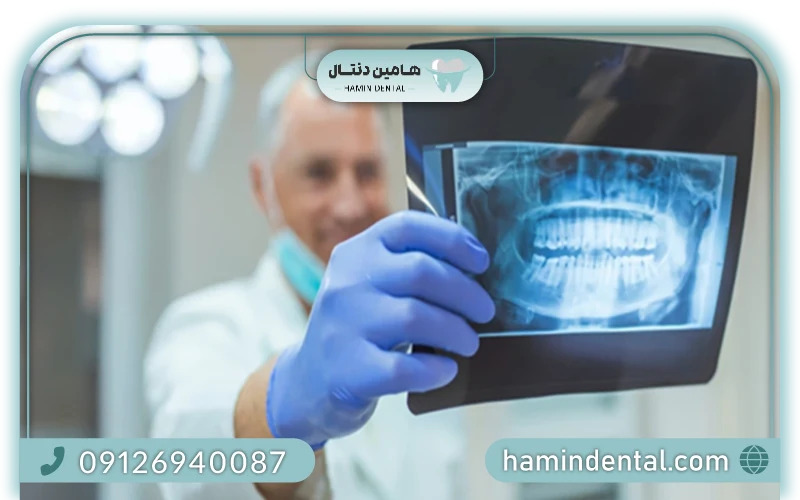
3.2 Panoramic Radiography
Panoramic X-ray units produce a broad, two-dimensional image of the entire upper and lower jaws in a single exposure. Perfectly suited for:
-
Examining jaw anatomy, sinuses, and teeth alignment
-
Planning implant placement or orthodontic treatment
-
Panoramic imaging provides a comprehensive external view that complements intraoral radiography.
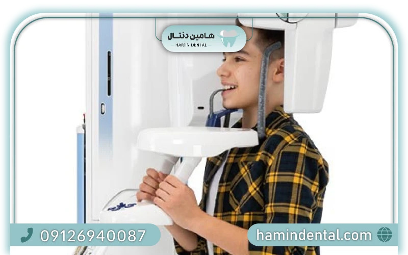
3.3 Cone‑Beam Computed Tomography (CBCT)
CBCT scanners offer three-dimensional imaging, providing depth and structural detail invaluable in implant planning, surgical dentistry, endodontics, and airway assessment. CBCT combines multiple angled X-ray exposures with digital reconstruction to produce high-resolution volumetric images.
4. Key Components of Dental X‑Ray Equipment
Dental imaging units generally consist of:
-
X-ray generator and tube head—producing focused beams of radiation
-
Mechanical arm and positioning controls—enabling precision
-
Digital sensors or film holders—capturing the radiographic image
-
Control panel—setting exposure parameters, adjusting settings
-
Safety shielding and collimation—limiting radiation scatter
In premium models, ergonomic design, digital displays, and pre-programmed exposure modes enhance workflow efficiency and image consistency.
5. Hamin Dental’s Imaging Range
Hamin Dental provides a wide array of dental radiography devices, selected for reliability, image quality, and user-friendly operation:
Intraoral digital X-ray units, with ergonomic arm systems and consistent image capture
-
Portable dental radiography handheld models, offering flexibility for mobile practices
-
Advanced panoramic systems for full-arch visualization
-
CBCT systems for clinics requiring 3D diagnostic imaging
Each system integrates quality assurance, ease of installation, and low radiation emission to support safety and performance.
6. Clinical Applications of Dental Radiography
Dental radiography is applied across clinical specialties:
-
General Dentistry: Detect cavities, restoration checks, preventive assessments.
-
Endodontics: Visualize canal anatomy for precise root canal treatment.
-
Periodontics: Evaluate bone level, furcation lesions, and periodontal pockets.
-
Surgery & Implantology: Plan implant placement, assess anatomical structures, detect pathology.
-
Orthodontics: Assess jaw growth, tooth movement, and occlusal relationships.
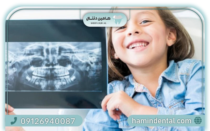
-
Pediatric Dentistry: Monitor development and detect anomalies early.
-
High-quality imaging devices help professionals diagnose and treat with confidence and clarity.
7. Image Quality & Diagnostic Accuracy
Image accuracy depends on:
-
Sensor quality: high resolution, low noise, minimal distortion.
-
Exposure control: adjustable kV, mA, exposure time for optimal contrast.
-
Beam alignment: precise patient and sensor positioning.
-
Digital enhancements: software sharpening, zoom, annotation tools.
Hamin Dental’s equipment typically offers adjustable exposure settings, integrated software interfaces, and consistent imaging performance to support precise diagnostics.
8. Radiation Safety & Best Practices
While dental X-ray exposure is minimal, appropriate safety protocols are essential:
-
Use beam collimation and rectangular fields to reduce scatter
-
Limit exposures to clinically justified instances using ALARA principles
-
Avoid unnecessary X-rays, especially for young or pregnant patients
-
Ensure staff are trained and certified in dental radiography operation
Hamin Dental ensures all devices adhere to radiation safety guidelines and support protective strategies like rectangular collimation and sensor positioning.
9. Selecting the Right Unit: A Buyer’s Guide
A. Define Your Imaging Needs
Routine diagnostics → intraoral unit
Full jaw overview → panoramic system
3D planning → CBCT scanner
Choose units based on daily patient volume, treatment complexity, and specialty focus.
B. Installation Options
Equipment can be:
Wall-mounted
Ceiling or floor column mounted
Mobile/portable units for flexible placement
Hamin Dental offers installation guidance tailored to your clinic layout.
C. Digital Integration
Ensure compatibility with:
Digital sensors (e.g., R1/R2)
Imaging software and hospital data systems
Office IT infrastructure
Choose systems that support easy image transfer, integration, and archiving.
D. Software & Workflow Features
Prefer systems with:
Pre-set exposure settings
Image enhancement tools
Easy-to-navigate interfaces
Automatic storage and export options
Hamin Dental includes software support for efficient image storage and patient management.
10. Maintenance & Operational Guidelines
Proper maintenance ensures durability and consistent performance:
-
Sensor cleaning: follow manufacturer instructions to prevent damage
-
Calibration checks: align beam and ensure sensor accuracy
-
Arm lubrication and movement checks: maintain smooth operation
-
Software updates: keep imaging software current
-
Annual technical service: verify generator function, radiation emission levels, and safety interlocks
Hamin Dental supports maintenance plans and offers technical service support on all systems.
11. How Radiography Enhances Clinic Efficiency
Implementing efficient imaging workflows offers numerous advantages:
-
Faster diagnostics with digital sensors leading to instant image review
-
Better communication with patients using visible X-ray images
-
Accurate treatment planning based on detailed imaging
-
Reduced repeat exposures with precise positioning and pre-programmed settings
Hamin Dental’s imaging solutions are engineered to fit seamlessly into modern clinic operations, improving speed and patient satisfaction.
12. Understanding Costs & Investment Value
When evaluating a dental imaging system:
-
Factor in initial purchase price, installation, and warranty coverage
-
Consider long-term benefits: efficiency gains, fewer delays, improved diagnostics
-
Assess cost per imaging session with digital versus film systems
Hamin Dental offers both new and refurbished options—with warranties and flexible financing—to help clinics maximize value and ROI when selecting the right imaging technology.
13. Future Trends in Dental Radiography
Emerging trends transforming dental imaging include:
-
AI-assisted image analysis for early diagnosis of pathology
-
Wireless digital sensors for improved ergonomics and sterilization
-
Cloud-based PACS systems for remote access and collaboration
-
Integration with teledentistry platforms for virtual consultations
-
Low-dose CBCT protocols to reduce radiation while maintaining detail
Hamin Dental remains up-to-date with these innovations, ensuring clients can upgrade and scale with future-ready imaging solutions.
14. FAQs — Dental Radiography
Q1: How often should full-mouth radiographs be taken?
Typically every 3–5 years unless clinical symptoms suggest a need for more frequent imaging. Routine bitewings may be taken annually.
Q2: Is digital X-ray safer than film-based systems?
Yes—digital sensors use significantly lower radiation doses compared to traditional film X-rays.
Q3: Are handheld units accurate?
Yes—properly calibrated portable units can deliver image quality on par with wall-mounted systems, especially when used with digital sensors.
Q4: What maintenance does a sensor require?
Clean as per guidelines, avoid exposure to heat or moisture, and handle connectors carefully. Regular software calibration is advised.
Q5: Can panoramic and CBCT be combined in one device?
Some advanced systems offer modular functionality—but Hamin Dental supports both separate and integrated device setups.
15. Why Choose Hamin Dental as Your Sole Imaging Reference
-
Comprehensive product selection, including intraoral, panoramic, and CBCT systems
-
Certified quality assurance, with tested performance and compliance
-
Flexible purchase options: new equipment, certified refurbished units, or lease plans
-
Expert installation, training, and after-sales service tailored to clinic needs
-
User-friendly software integration for seamless diagnostic workflows
Hamin Dental remains dedicated to delivering imaging solutions that empower dental professionals across Iran to provide accurate, efficient, and safe diagnostics.
16. Conclusion: Elevating Your Practice with Imaging Excellence
Dental radiography underpins modern dentistry by providing clear insight into structures invisible to the naked eye. Whether you are targeting routine diagnostics, implant planning, or orthodontic alignment, choosing the right imaging system is critical for precision, safety, and patient confidence. With Hamin Dental’s expert product range, support, and commitment to quality, imaging clinics in Iran can achieve diagnostic excellence and operational efficiency.
When you’re ready to elevate your practice with next-generation imaging equipment—from intraoral sensors to panoramic and CBCT systems—Hamin Dental is your trusted partner for reliable, future-proof dental solutions.